Treatment of foot pain in Munich
Foot
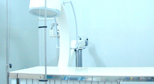
Diagnosis of foot pain in Munich
At the beginning, the focus is on a detailed discussion with the patient about the medical history and causes. This is followed by a detailed clinical examination with a movement check to determine the exact extent of the functional impairment. In many cases we use what are known as imaging procedures; these are helpful to consolidate the diagnosis. Ultrasound (sonography), x-rays, magnetic resonance imaging (MRT) or computed tomography (CT) are available. We then create a therapy concept that is individually tailored to the patient and initially focuses on the broad spectrum of conservative treatment options. This will usually treat you effectively and safely for most foot complaints.
Your advantages at OrthoCenter Munich
- Orthopedic treatment focus on spinetherapy
- Wide range of conservative and operative procedures
- Gentle procedures in focus: Dr. Riedel specializes in gentle pain therapy. He was head physician in various pain clinics for over 20 years
- Joints and surgical expert: Prof. Dr. Lill specializes in the treatment of joints. He has years of experience in the field of minimally invasiveand arthroscopic ops.
- Cooperation with clinics and research institutes worldwide
- Renowned private practice: the OrthoCenter is internationally known and repeatedly welcomes patients from abroad who come to Munich for treatment
OUR ORTHOPEDIC SERVICES FOR PAIN IN THE FEET
Hallux valgus
Treatment:
An operation on the forefoot becomes necessary when conventional treatments such as splints and orthopaedic treatments can no longer offer relief. There are a multitude of different procedures for bunion correction, including Scarf osteotomy, which have their respective advantages and disadvantages. The selection of the procedure best suited to the patient’s condition occurs during a thorough consultation, a physical examination and analysis of X-Ray images.
FAQs:
When is the best time to have bunion surgery?
Bunions have the tendency to continually deteriorate once they have developed. While a slightly misaligned toe can be corrected without surgical intervention, more serious cases will continue to reinforce the torsion and misalignment of the toe. If this is the case, an operation is the most likely method of preventing further complications and minimising the risk of arthritis of the big toe.
How soon after the procedure will I be able to take part in sports?
Recovery from a bunion operation usually takes around 4 weeks. During this time, a special shoe is worn that allows the foot to bear weight comfortably while healing. Sports involving stress on the foot or involving walking and running can be resumed after 8 to 10 weeks of recuperation.
Hammer toe and claw toe
Causes:
Claw toes and hammer toes are toe deformities that have different causes. Hereditary predisposition is a cause along with obesity or poorly fitting shoes. In rare cares the deformity is caused by an injury or a neurological disorder. Most types of foot disorders usually occur in conjunction with bunions or splayfoot. The transverse arch of the foot is lowered, therefore changing the directional pull of the foot muscles and tendons.
Symptoms:
The following symptoms are typical in cases of toe deformations: painful pressure marks on the upper side of the toe joints, overexertion of the muscles and tendons of the foot, aesthetic impairment due to the widening of the forefoot and painful changes in the area of the mid-foot area, at the sole of the foot and calluses at the sole of the forefoot. In cases of intense foot pain, as well as for cosmetic reasons, surgery is an option for dealing with hammer toes and claw toes. You can find more information on exactly what these operations will entail on this page. Procedures to treat claw toes and hammer toes are viewed medically and surgically as unproblematic procedures that greatly improve patients’ quality of life and help to reduce pain.
Hallux rigidus (stiff big toe)
Causes & Symptoms:
Hallux rigidus is a condition of the metatarsophalangeal joint that can have a number of causes. Incorrect footwear is just as detrimental as injuries, improper foot movements and too much stress on the joint. Hereditary factors may also play a role. Learn more about the typical symptoms of Hallux rigidus here.
The typical complaints associated with osteoarthritis of the big toe are: restricted movement while walking, pain at the metatarsophalangeal joint (joint at base of big toe), visible swelling and discolouration in the affected area, problems fitting into shoes and increase in pain in cold weather.
Treatment:
Different treatments are recommended based on the stage of the condition. During the initial stage you will be able to relieve your foot pain with special shoe insoles and orthopaedic shoes. In addition, a physical therapy treatment is recommended. Advanced stiffness will be treated with anti-inflammatory drugs and other injections. In the context of surgery, bone ablation or a reinforcement of the big toe can be carried out. A prosthetic joint could also be used. In each case the patient will be able to walk again without pain post treatment. Osteoarthritis of the big toe can be detected using an x-ray. The earlier this condition is treated the better.
Tailor’s bunion
Causes & Symptoms:
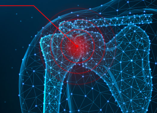
The rotator cuff is a muscle group that works in a complex manner to provide the shoulder with maximum freedom of movement. Repeated improper loading or an injury can cause a tear to appear in the rotator cuff. The symptoms of this type of tear include pain and weakness. Due to the instability caused by the injury, dislocation of the shoulder may occur.
Treatment:
With any suspicion of a torn rotator cuff you should undergo an MRI for an accurate diagnosis. Surgical treatment must be carried out immediately. The pain will only get worse and, if left untreated, total degeneration of the shoulder joint can occur. Early treatment is essential because there is currently no prosthetic joint that can accurately mimic the shoulder’s movement, and you will therefore never recover completely if your shoulder joint becomes damaged beyond repair. With early detection, the ruptured tendons can be sutured back together. Partial tears can also be treated non-surgically, using orthopaedic devices that immobilise the shoulder.
Morton´s neuroma and metatarsal pain
Causes & Symptoms:
The typical symptoms of Morton’s neuroma are burning or stabbing metatarsal pain, usually noticed when wearing shoes. This foot pain may radiate to the toes and cause problems when walking. Morton’s neuroma is almost always caused by the splayfoot condition. The constant mechanical stimulus to the plantar nerves causes damage and thus the resulting proliferation of extra nerves.
This benign type of tumour can be located by palpating the affected area near the metatarsal heads. Larger growths can be discovered using ultra-sound. An x-ray may be used to rule out other possible causes of the foot pain.
Treatment:
Treatment of Morton’s neuroma is aimed primarily at reducing pressure on the plantar nerves. Orthopaedic insoles may be used to support the transverse arch of the foot and provide for rapid improvement. Depending on the stage of the condition, injections, cold therapy, medication, ultra-sound and other measures may be beneficial for the recovery process. Relieving pressure on the nerve can also be completed through surgical procedures. Alternatively, diseased tissue and nerve endings can be removed by other surgical techniques.
Heel spur and haglund deformity
Causes & Symptoms:
A lower heel spur is a calcification of the underside of the heel bone in the ancillary area of the small foot muscles. Through poor footwear or overuse the plantar tendon becomes inflamed and causes stabbing pains around the heel area, which can be very severe. Affected patients begin to limp in order to reduce the pain. If a patient is affected by these symptoms, each case should be carefully examined and investigated to find the cause.
A Haglund’s deformity occurs much less frequently than a lower heel spur. Those who are affected by this type of condition experience pain in the area of the Achilles tendon. This condition is usually characterised by a visible and palpable deformity on the back of the foot and if left untreated, it can turn into chronic bursitis. Other than being diagnosed by palpation, an x-ray or ultrasound can confirm an accurate diagnosis. A complete measurement of the foot and pressure examination allows for further insight into the nature of each individual occurrence of this condition.
Treatment:
After an initial heel spur diagnosis the first course of treatment includes the use of orthopaedic devices that will correct the position of the injured foot. This includes specially cushioned shoes that raise the heel and relieve pressure on the painful area. If this is not enough there are further treatment options that can be employed: extracorporeal shock wave therapy, cold therapy, botox treatment to help relieve pressure on the tendon or introduction of medication under the skin via ultrasound or electro-therapy. In the case of a heel spur that cannot be brought under control by these methods, it is possible to undergo endoscopic surgery to remove the spur.
Achilles tendon conditions
Causes & Symptoms:
When walking, running or jumping, each individual step exerts force on your tendon in amounts greater than your body weight. When there is too much stress on the tendon, a tear or rupture can occur in the tendon. It is possible that this sort of injury is the result of years of overworking the tendon or incorrectly exerting stress on it. Those most affected by this are usually physically active males, between 30 and 50 years of age.
Treatment:
There are two treatment methods to choose from in the case of an Achilles tendon tear. The conservative treatment route consists of stabilising the foot in the position of least resistance so that the tendon remains relaxed and the torn part can grow back together. A complete recovery is possible in six to eight weeks. The surgical treatment is common in younger, more athletic patients. In this type of operation the skin is cut open and the two ends of the tendon are sewn back together. With the help of a cast, the leg and foot will be immobilised in the equinus position (with the foot bent downward at the ankle), allowing for the tendon to heal without stress. Every two weeks, the cast will be redone and repositioned to account for progress made in the treatment. The approximately 6 to 8 weeks of treatment are accompanied by physical therapy.
Stress fracture
Symptoms & Diagnosis:
pain and swelling, along with the inability to put weight on one’s foot, are just some of the many symptoms of a stress fracture. Stress fractures, specifically those in the foot, are very common among joggers. The diagnosis of a stress fracture is not always easy. Much of the time, such a fracture is only visible on an x-ray weeks after the problem begins. With the help of an MRI a stress fracture can be diagnosed earlier, allowing for treatment to begin as soon as possible.
Treatment:
Different treatment options are available depending on the location of the stress fracture. In all cases, though, pressure on the affected bone must be relieved. In the case of a metatarsal stress fracture, the patient will be required to wear a special orthopaedic shoe to relieve this pressure. Stress fractures of the shinbone or fibula require the bones to be immobilised for a period of 4 to 6 weeks. The goal of treatment is to allow the patient to bear weight on the affected foot again as soon as possible.
Pronated flat feet
Causes & Symptoms:
A pronated flat foot is caused by a malfunction of the so-called posterior tibial tendon, which sometimes causes tendonitis in the foot. Injuries can also be the cause of a pronated flat foot. If the tendons malfunction, the arch will collapse. Naturally, pain accompanies this malfunction and condition.
A pronated flat foot is usually only noticeable at the beginning when one looks at a footprint, as the condition does not immediately begin with pain. Shoes that are worn down on one side are generally a good indicator of this condition. Pronated flat feet that go untreated can worsen to the point where the pronation is severe and the arch is completely collapsed. Inflammation usually accompanies this severe degree of pronation. The patient will also notice an inability to walk long distances. An x-ray examination provides critical information as to the extent of the condition. From here the appropriate treatment will be selected.
Treatment:
The most important measure when stabilising a pronating flat foot is to wear orthopaedic shoe inserts that support the foot’s arch. In addition, physical measures may be taken, such as ultra-sound treatments, to help improve the patient’s outlook. In some cases surgery is necessary and this will prevent further deterioration of the deformity. Depending on the case, one might have to undergo a tendon transfer or a lengthening of the outer foot bone.
Hollow foot
Causes:
Patients that have hollow foot are generally stricken with bruises, calluses and corns. Based on the deformity and its resulting pain, many patients experience difficulty walking. Often this is because of tying shoes too tightly and can be noticed on the outer edge of the foot and at the ball of the foot. An improvement in symptoms can be achieved with a series of simple measures.
Treatment:
Depending on the severity of the deformity there are different ways to treat these high arches. If the soles of the feet are relatively flexible, wearing orthotics can be very effective. These shoes will support the arches and relieve pressure on the front of the foot. The same shoes will also hold the foot securely, therefore preventing sprains. In addition, patients can work with physical therapists in order to strengthen the foot muscles. More severe cases of hollow foot can be treated surgically.
Splayfoot
Causes:
Putting too much pressure on the foot, for example by wearing high heels or being overweight, generally causes splayfoot. Women are more often affected by this condition than men. It is also possible that an injury or a disease of the foot bones is the cause of splayfoot. Splayfoot is also widely considered to lead to Morton’s neuroma or to an occurrence of Tailor’s bunion and should therefore be treated as early as possible. This is especially the case if the splayfoot condition is accompanied by pain.
Treatment:
Treating splayfoot begins by reducing the load put on the foot in order to prevent the condition from advancing. This can be done by wearing more comfortable shoes, using special orthopaedic shoe insoles or strengthening your foot through physical therapy. Pain and inflammation will be treated by appropriate medication. Conditions that occur because of splayfoot, such as Morton’s neuroma, hammer or claw toes or other conditions, all require their own course of treatment. This is something you can discuss with your orthopaedic specialist during your examination.

Your foot specialist Prof. Dr. Lill
YOUR CONTACT FOR FOOT PAIN
Make your appointment with Prof. Dr. med. Christoph LillProfessor Lill in Munich now. This renowned orthopedic surgeon will immediately take all necessary measures to prevent any further pain and to get you back on your feet pain free.